Article courtesy of: Review of Ophthalmology, Published 30 December 2005
Management Options for Macular Edema
Many strategies have been used to manage macular edema, with varying degrees of success.
“Macular edema represents the pathologic accumulation of extracellular fluid within the retina, primarily in the outer plexiform and inner nuclear layers, as a nonspecific response to a breakdown in the blood-retinal barriers. ME is a frequent cause of vision loss in patients with diabetes mellitus, retinal venous occlusion, uveitis, and following intraocular surgery. It occurs less frequently in the setting of vitreoretinal traction, choroidal neovascularization and a number of other conditions. Many strategies have been employed to manage ME with varying success. This article reviews the available treatment options for this common condition” (Ober, Klais, & Cunningham, Jr., 2005)
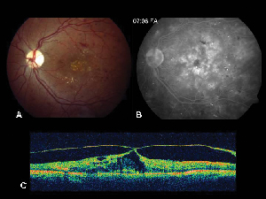
Figure 2. A. Color fundus photograph of the left eye of a patient with non-proliferative diabetic retinopathy and lipid exudation in and around the fovea. B. Late-phase FA reveals macular edema in the central macula. C. Optical coherence tomography demonstrates the abnormal vitreoretinal interface as well as macular edema.
Images courtesy of Ophthalmology Review Journal.

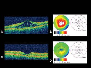
| Figure 4. A. Optical coherence tomography image of an eye with diabetic macular edema with corresponding retinal thickness mapB. generated by the OCT. C. OCT of the same patient one month following intravitreal triamcinolone acetonide injection withcorresponding retinal thickness map. D. Resolution of macular edema. Visual acuity improved from 20/200 to 20/80 following treatment. |
Read the entire article by clicking here.
Reference
Ober, M., D. MD., Klais, C., M. MD., & Cunningham, Jr., E., T. MD. (2005). Management Options for Macular Edema. Review of Ophthalmology. Retrieved from https://www.reviewofophthalmology.com/article/management-options-for-macular-edema


